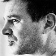
Kenneth L Jacobs
age ~45
from Royersford, PA
- Also known as:
-
- Kenneth Lloyd Jacobs
- Kent L Jacobs
- Ken Jacobs
Kenneth Jacobs Phones & Addresses
- Royersford, PA
- Audubon, PA
- Winterville, NC
- Tulsa, OK
- Media, PA
- 1023 Thrush Ln, Norristown, PA 19403 • 6107504999
Work
-
Position:Machine Operators, Assemblers, and Inspectors Occupations
Education
-
School / High School:University Of North Carolina, Chapel Hill, School Of Medicine1992
Languages
English
Specialities
Obstetrics & Gynecology • Clinical Pathology
Lawyers & Attorneys

Kenneth Jacobs - Lawyer
view sourceOffice:
Kenneth S. Jacobs
Specialties:
Estate Planning
Probate Law
Landlord and Tenant Law
Elder Law
Trusts & Estates
Wills & Probate
Trusts
Probate Law
Landlord and Tenant Law
Elder Law
Trusts & Estates
Wills & Probate
Trusts
ISLN:
900955950
Admitted:
1991
University:
University of California, Berkeley, B.A., 1988
Law School:
University of Washington, J.D., 1991

Kenneth Jacobs - Lawyer
view sourceOffice:
K. P. Jacobs & Associates, P.L.C.
Specialties:
Communications & Media
Communications & Media
Entertainment
Entertainment
General Practice
Communications & Media
Entertainment
Entertainment
General Practice
ISLN:
914706937
Admitted:
1995

Kenneth Jacobs - Lawyer
view sourceOffice:
GrayRobinson, P.A.
Specialties:
Banking & Finance
Bankruptcy & Creditors' Rights
Business Law
Employment & Labor
Labor and Employment Law
Title Insurance and Real Estate Litigation
Bankruptcy & Creditors' Rights
Business Law
Employment & Labor
Labor and Employment Law
Title Insurance and Real Estate Litigation
ISLN:
901304061
Admitted:
1992
University:
Duke University, B.A., 1989
Law School:
University of Florida College of Law, J.D., 1992

Kenneth Jacobs - Lawyer
view sourceOffice:
Smith Buss & Jacobs, LLP
Specialties:
Business
Contracts & Agreements
Mortgage Financing
Sales and Acquisitions
Syndication or Transactional Real Estate
Contracts & Agreements
Mortgage Financing
Sales and Acquisitions
Syndication or Transactional Real Estate
ISLN:
906220427
Admitted:
1979
University:
Wesleyan University, B.A., 1974
Law School:
New York University, J.D., 1978
Name / Title
Company / Classification
Phones & Addresses
Director, President
Lazard Freres & Co. LLC
KENNETH JACOBS, D.O., LLC
JACOBS & SONS LOGGING, LTD
JACOBS FLOOR COVERINGS, LLC
Resumes

Kenneth Jacobs
view source
Kenneth Jacobs
view source
Kenneth Jacobs
view source
Kenneth Jacobs
view source
Kenneth Jacobs
view source
Visiting Faculty Member, Department Of Architecture, Philadelphia University
view sourceLocation:
Greater Philadelphia Area
Industry:
Architecture & Planning
Isbn (Books And Publications)


The Emerging Universe: Essays on Contemporary Astronomy
view sourceAuthor
Kenneth C. Jacobs
ISBN #
0813903971

License Records
Kenneth C Jacobs
License #:
143 - Expired
Category:
Health Care
Issued Date:
Sep 20, 1976
Effective Date:
Jan 1, 1901
Expiration Date:
Jan 31, 1993
Type:
Occupational Therapist
Medicine Doctors

Dr. Kenneth L Jacobs - MD (Doctor of Medicine)
view sourceSpecialties:
Obstetrics & Gynecology
Clinical Pathology
Clinical Pathology
Languages:
English
Education:
Medical School
University Of North Carolina, Chapel Hill, School Of Medicine
Graduated: 1992
University Of North Carolina, Chapel Hill, School Of Medicine
Graduated: 1992

Kenneth M. Jacobs
view sourceSpecialties:
Pulmonary Disease
Education:
Medical School
SUNY Downstate Medical Center College of Medicine
Graduated: 1976
SUNY Downstate Medical Center College of Medicine
Graduated: 1976
Procedures:
Lung Biopsy
Allergy Testing
Pulmonary Function Tests
Allergy Testing
Pulmonary Function Tests
Conditions:
Bronchial Asthma
Pneumonia
Pulmonary Embolism
Acute Bronchitis
Cardiac Arrhythmia
Pneumonia
Pulmonary Embolism
Acute Bronchitis
Cardiac Arrhythmia
Description:
Dr. Jacobs graduated from the SUNY Downstate Medical Center College of Medicine in 1976. He works in Scranton, PA and specializes in Pulmonary Disease. Dr. Jacobs is affiliated with Regional Hospital Of Scranton.

Kenneth Jacobs
view sourceSpecialties:
Internal Medicine
Pulmonary Disease
Critical Care Medicine
Critical Care Medicine
Pulmonary Disease
Critical Care Medicine
Critical Care Medicine
Education:
State University of New York Downstate (1976)
Wikipedia References

Kenneth M . Jacobs
Work:
Position:
Businessman • American chief executive • Chairman • CEO
Education:
Studied at:
Stanford Graduate School of Business
Us Patents
-
Flow Cytometer Apparatus For Three Dimensional Difraction Imaging And Related Methods
view source -
US Patent:20110090500, Apr 21, 2011
-
Filed:Jun 11, 2009
-
Appl. No.:12/997186
-
Inventors:Xin-Hua Hu - Greenville NC, US
Kenneth M. Jacobs - Greenville NC, US
Jun O. Lu - Greenville NC, US -
International Classification:G01N 21/53
-
US Classification:356337
-
Abstract:A flow cytometer assembly includes a fluid controller configured to form a hydrodynamically focused flow stream including an outer sheath fluid and an inner core fluid. A coherent light source is configured to illuminate a particle in the inner core fluid. A detector is configured to detect a spatially coherent distribution of elastically scattered light from the particle excited by the coherent light source. An analyzing module configured to extract a three-dimensional morphology parameter of the particle from a spatially coherent distribution of the elastically scattered light.
-
Near-Infrared Fluorescence Imaging For Blood Flow And Perfusion Visualization And Related Systems And Computer Program Products
view source -
US Patent:20200305721, Oct 1, 2020
-
Filed:Mar 25, 2020
-
Appl. No.:16/829468
-
Inventors:- Greenville NC, US
JIAHONG JIN - Greenville NC, US
KENNETH MICHAEL JACOBS - Greenville NC, US
TAYLOR FORBES - Greenville NC, US
BRYENT TUCKER - Rocky Mount NC, US
XIN HUA HU - Greenville NC, US -
International Classification:A61B 5/00
G06T 5/00
G01N 21/64 -
Abstract:Systems for obtaining an image of a target are provided including at least one multi-wavelength illumination module configured to illuminate a target using two or more different wavelengths, each penetrating the target at different depths; a multi-wavelength camera configured to detect the two or more different wavelengths illuminating the target on corresponding different channels and acquire corresponding images of the target based on the detected two or more different wavelengths illuminating the target; a control module configured synchronize illumination of the target by the at least one multi-wavelength illumination module and detection of the two or more different wavelengths by the camera; an analysis module configured to receive the acquired images of the target and analyze the acquired images to provide analysis results; and an image visualization module modify the acquired images based on the analysis results to provide a final improved image in real-time.
-
A Laser Safety Adaptor For Use In Laser Based Imaging Systems And Related Devices
view source -
US Patent:20180337507, Nov 22, 2018
-
Filed:Mar 24, 2016
-
Appl. No.:15/559646
-
Inventors:- Greenville NC, US
Cheng Chen - Greenville NC, US
Kenneth Michael Jacobs - Greenville NC, US -
International Classification:H01S 3/00
G02B 27/30
G02B 5/02
G02B 27/09
G02B 6/28 -
Abstract:A fiber assembly is provided including a laser input end configured to receive an input signal having a first laser beam intensity. The fiber assembly further includes a plurality of channels attached to the laser input end and a plurality of laser safety adaptors. Each of the plurality of laser safety adaptors is configured to receive a corresponding one of the plurality of channels. A laser beam exiting each of the plurality of laser safety adaptors has a second laser beam intensity that is less than the first laser beam intensity.
-
Multi-Wavelength Beam Splitting Systems For Simultaneous Imaging Of A Distant Object In Two Or More Spectral Channels Using A Single Camera
view source -
US Patent:20180067327, Mar 8, 2018
-
Filed:Mar 22, 2016
-
Appl. No.:15/559605
-
Inventors:- Greenville NC, US
Cheng Chen - Greenville NC, US
Kenneth Michael Jacobs - Greenville NC, US -
International Classification:G02B 27/10
G02B 27/14
G03B 33/12
H04N 9/09
G02B 27/00 -
Abstract:An optical imaging system and related methods are provided that acquire images of an object at a distance in different spectral regions using only one camera. The systems and methods are adaptable to applications where information (simultaneous or sequential) from more than one spectral region is of interest while only one camera is available or entailed.
-
Multi-Spectral Physiologic Visualization (Mspv) Using Laser Imaging Methods And Systems For Blood Flow And Perfusion Imaging And Quantification In An Endoscopic Design
view source -
US Patent:20180020932, Jan 25, 2018
-
Filed:Aug 28, 2017
-
Appl. No.:15/688472
-
Inventors:- Greenville NC, US
Kenneth Michael Jacobs - Greenville NC, US -
International Classification:A61B 5/026
A61B 1/06
A61B 1/00
A61B 1/005
A61B 5/00
A61B 1/04
A61B 1/313 -
Abstract:Multispectral imaging systems are provided including a first light source having a first wavelength configured to image a sample; a second light source, different from the first light source, having a second wavelength, different from the first wavelength, configured to image the sample; and at least a third light source, different from the first and second light sources, having a third wavelength, different from the first and second wavelengths, configured to image the sample. A camera is configured to receive information related to the first, second and at least third light sources from the sample. A processor is configured to combine the information related to the first, second and at least third light sources provided by the camera to image an anatomical structure of the sample, image physiology of blood flow and perfusion of the sample and/or synthesize the anatomical structure and the physiology of blood flow and perfusion of the sample in terms of a blood flow rate distribution. The imaging system is directed and focused on a field of view (FOV) in a region of interest of the sample using an endoscope.
-
Methods, Systems And Computer Program Products For Determining Hemodynamic Status Parameters Using Signals Derived From Multispectral Blood Flow And Perfusion Imaging
view source -
US Patent:20170274205, Sep 28, 2017
-
Filed:Oct 13, 2015
-
Appl. No.:15/518545
-
Inventors:- Greenville NC, US
Sunghan Kim - Winterville NC, US
Zhiyong Peng - Greenville NC, US
Kenneth Michael Jacobs - Greenville NC, US -
International Classification:A61N 1/08
G06F 19/00
G06T 7/00
A61B 5/05 -
Abstract:Methods for calculating a MetaKG signal are provided. The method including illuminating a region of interest in a sample with a near-infrared (NIR) light source and/or a visible light source; acquiring images of the region of interest; processing the acquired images to obtain metadata associated with the acquired images; and calculating the MetaKG signal from the metadata associated with the acquired images. Related systems and computer program products are also provided.
-
Methods, Systems And Computer Program Products For Visualizing Anatomical Structures And Blood Flow And Perfusion Physiology Using Imaging Techniques
view source -
US Patent:20170224274, Aug 10, 2017
-
Filed:Oct 13, 2015
-
Appl. No.:15/518548
-
Inventors:- Greenville NC, US
Kenneth Michael Jacobs - Greenville NC, US
Zhiyong Peng - Greenville NC, US -
International Classification:A61B 5/00
A61B 5/0275
G06T 7/00
A61B 5/026 -
Abstract:Methods for combining anatomical data and physiological data on a single image are provided. The methods include obtaining an image, for example, a raw near-infrared (NIR) image or a visible image, of a sample. The image of the sample includes anatomical structure of the sample. A physiologic map of blood flow and perfusion of the sample is obtained. The anatomical structure of the image and the physiologic map of the sample are combined into a single image of the sample. The single image of the sample displays anatomy and physiology of the sample in the single image in real time. Related systems and computer program products are also provided.
-
Multi-Spectral Laser Imaging (Msli) Methods And Systems For Blood Flow And Perfusion Imaging And Quantification
view source -
US Patent:20160270672, Sep 22, 2016
-
Filed:Feb 26, 2016
-
Appl. No.:15/054830
-
Inventors:- Greenville NC, US
Zhiyong Peng - Greenville NC, US
Kenneth Michael Jacobs - Greenville NC, US -
International Classification:A61B 5/026
A61B 5/00 -
Abstract:Some embodiments of the present inventive concept provide a system that uses two wavelengths of differential transmittance through a sample to apply laser speckle or laser Doppler imaging. A first of the two wavelengths is within the visible range that has zero or very shallow penetration. This wavelength captures the anatomical structure of tissue/organ surface and serves as a position marker of the sample but not the subsurface movement of blood flow and perfusion. A second wavelength is in the near Infra-Red (NIR) range, which has much deeper penetration. This wavelength reveals the underlying blood flow physiology and correlates both to the motion of the sample and also the movement of blood flow and perfusion. Thus, true motion of blood flow and perfusion can be derived from the NIR imaging measurement without being affected by the motion artifact of the target.

Kenneth Jacobs
view source
Kenneth Jacobs
view source
Kenneth Jacobs
view source
Kenneth G Jacobs
view source
Kenneth Jacobs
view source
Kenneth R Jacobs Sr.
view source
Kenneth Debbie Jacobs
view source
Ken Jacobs
view sourceGoogleplus

Kenneth Jacobs
Work:
ConAgra Foods - Maintenance Planning Specialist (6)
US Navy - MM1N (5-5)
US Navy - MM1N (5-5)
Education:
Colorado Technical University Online - Network Management, Minico Senior High School
Tagline:
Still moving along

Kenneth Jacobs
Education:
Johnson & Wales University - Travel & Tourism
Tagline:
Kenny Jay

Kenneth Jacobs
Tagline:
I'm single im probably will stay single for the rest of my life and is a Choice.

Kenneth Jacobs
Bragging Rights:
Swiss

Kenneth Jacobs

Kenneth Jacobs

Kenneth Jacobs

Kenneth Jacobs
Flickr
Plaxo

Kenneth Jacobs
view sourceGlendale, CACEO at Lewis Howard & Assoc LLC Past: Regional Credit Manager at Rochux International
Myspace
Classmates

Kenneth Jacobs
view sourceSchools:
Walbrook High School Baltimore MD 1977-1981
Community:
Christina Daniels, Denise Harrid, Valerie Brown

Kenneth Jacobs
view sourceSchools:
Washington High School Brainerd MN 1953-1957
Community:
Everett Nelson, Steven White, Donna Schrom

Kenneth Logan (Jacobs)
view sourceSchools:
Roosevelt High School San Antonio TX 1996-2000
Community:
Robert King, Edmund Williams, Paul Dockery, James Wright

Kenneth Jacobs
view sourceSchools:
University School Milwaukee WI 1973-1977
Community:
Maree Deni, Robert Tarpey, Jim Wood, R Hafemann, Jamesc Jim

Kenneth Jacobs
view sourceSchools:
Maclay Middle School Pacoima CA 1966-1970
Community:
Rudy Serrano, Donna Dahms

Kenneth Jacobs
view sourceSchools:
Lunenburg High School Lunenburg MA 1976-1980
Community:
William Antoniac

Kenneth Jacobs
view sourceSchools:
Mansfield High School Mansfield PA 2004-2008
Community:
Michele Losinger

Kenneth Jacobs
view sourceSchools:
Chattahoochee High School Chattahoochee FL 2000-2004
Community:
Jerry Winters, Dennis Barber, James Howard, Nancy Cutchins
News

Civil Air Patrol thanks Capital Regional employees for work during COVID
view source- "They've done a lot to help all this community and help everybody," said Kenneth Jacobs, Captain Commander of the Civil Air Patrol Tallahassee 432."It's been a lot of hours through the nurses, therapists, labs, even down to the people that helped keep the hospital clean so that patients have a safe
- Date: Feb 15, 2021
- Category: More news
- Source: Google

Fed Ends QE While Keeping 'Considerable Time' Low-Rate Pledge
view source- S. chief executive officers see reasons for optimism. JPMorgan Chase & Co. CEO Jamie Dimon said last week that the worlds largest economy has no real weak spot, while Kenneth Jacobs, chief of investment bank Lazard Ltd., said on an earn ings call that the U.S. economy continues to be resilie
- Date: Oct 29, 2014
- Source: Google

Lazard Profit Falls 50% as Asset-Management Revenue Drops
view source- st monthdisclosed a 5.1 percent stake in Lazard, making it the biggestshareholder, according to data compiled by Bloomberg. The fundsaid Lazard, run by Chief Executive Officer Kenneth Jacobs, issignificantly undervalued and that earnings per share couldincrease to more than $3.50 in 2014.
- Date: Jul 26, 2012
- Category: Business
- Source: Google
Youtube
Get Report for Kenneth L Jacobs from Royersford, PA, age ~45


